

PaX-i Insight
衛部醫器輸字第030585號 PCH-30CS 本網站內容僅供牙醫師及牙醫診所專業從業人員參考使用,醫療諮詢請洽牙科醫師
醫師如需了解更詳細的產品資訊,歡迎與博泰業務團隊聯繫,我們將會用最熱誠的態度為您服務!

|
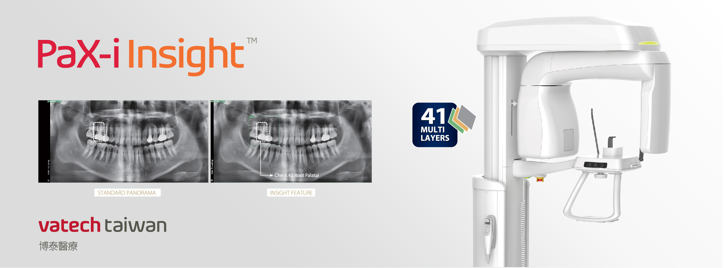
2D 新技術 Insight Pan
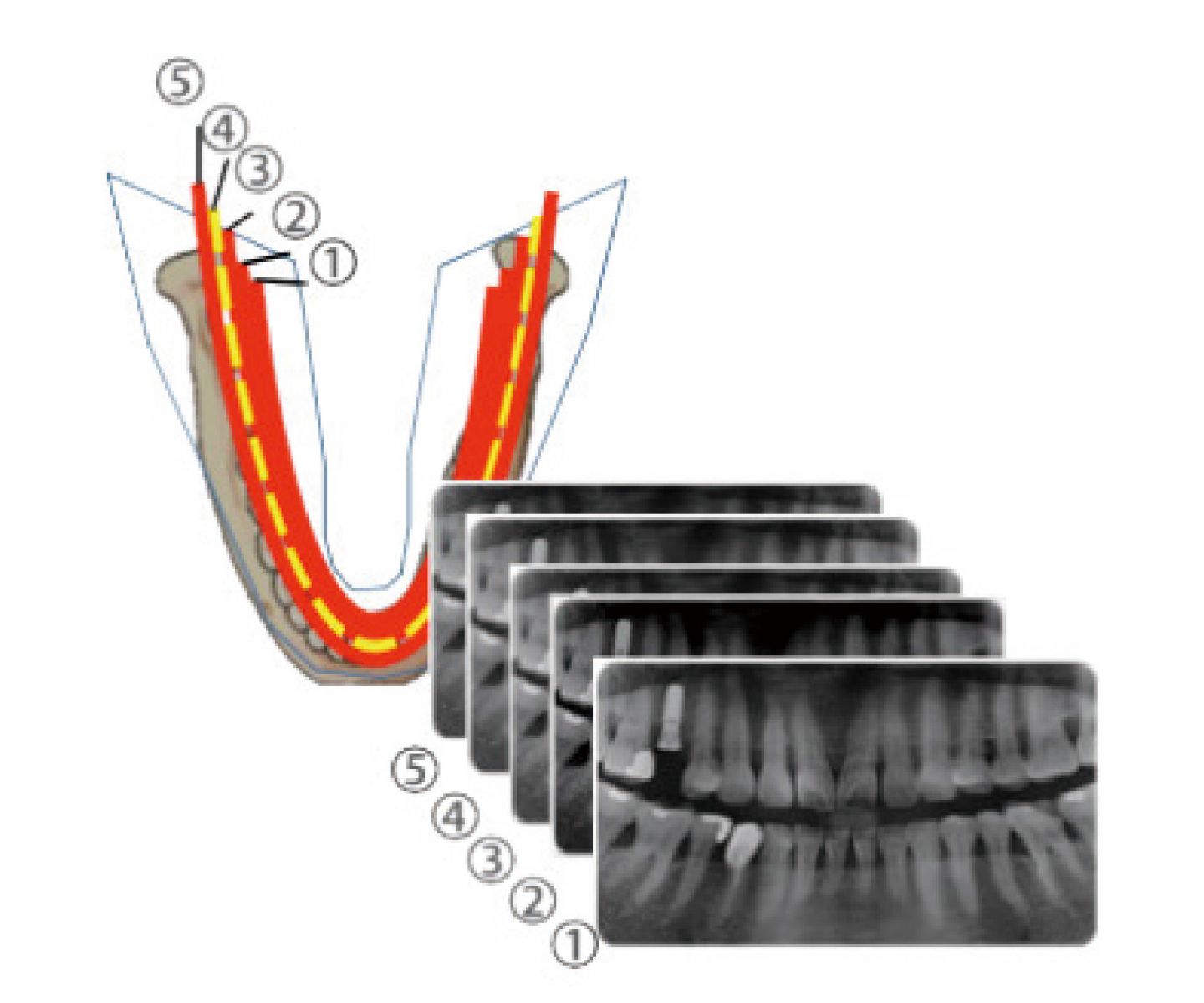
每位病患的牙弓尺寸各不相同,但傳統的 Panoramic 影像多以標準預設牙弓進行掃描產生一張 Panoramic 影像,因此容易造成前牙區產生不正確倍率的放大或縮小。 |
|
Insight Pan |
|
Insight Pan 於臨床上的優勢
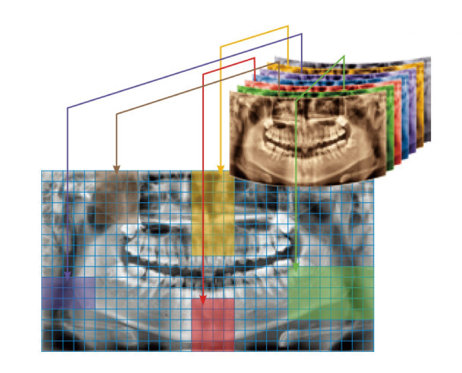
• 41 剖面清楚解析牙周顎側、頰側、舌側、唇側,讓醫師能進行更精準的判斷。
|
Innovation History
|
1 st Generation
One Layer |
2 nd Generation
Auto Focusing |
3 rd Generation
MagicPan |
 |
 |
 |
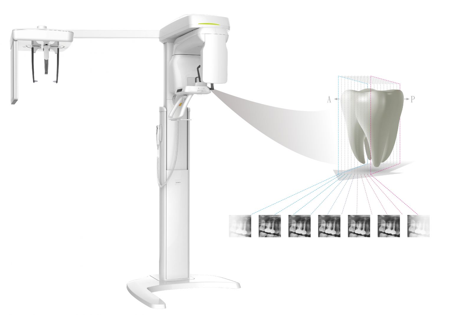
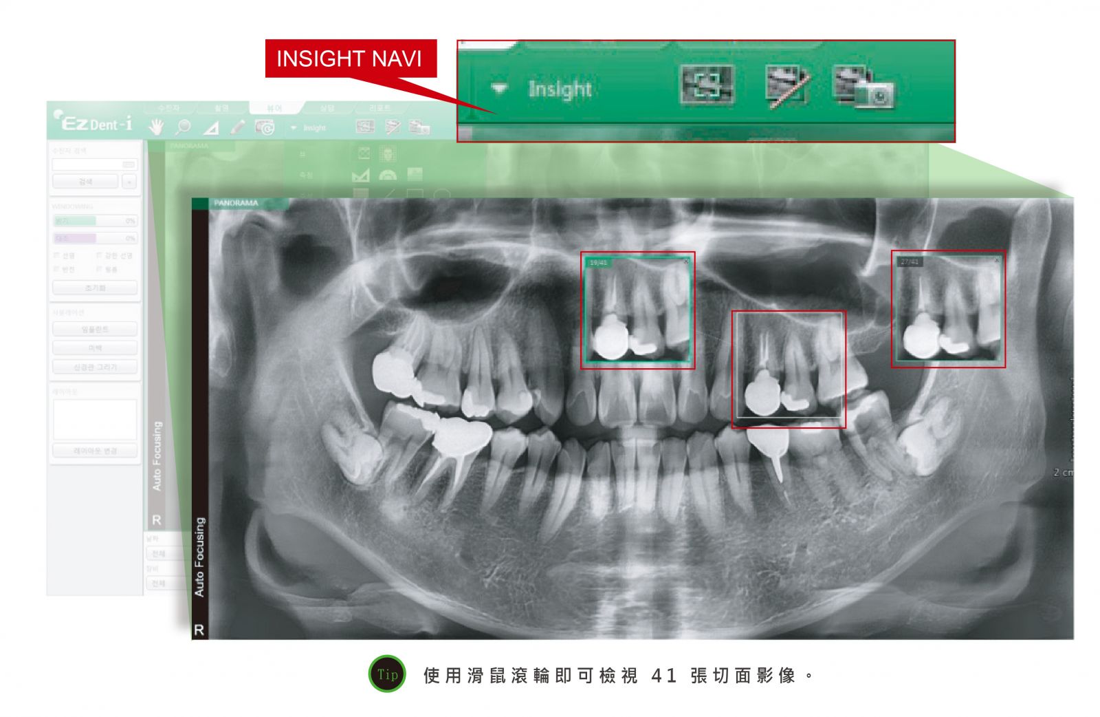
Insight Navi
|
專門軟體全方位檢視患者牙齒口腔資訊
為 PaX-i Insight 特別開發的 Insight Navi 功能,讓醫師在 EzDent-i 上開啟 Panoramic 影像後,點按牙位能借由滑鼠滾輪看到不同牙位的 Anterior 到 Posterior 影像,選定不同的切片,可探索更深入,如 Mesiobuccal, Distobuccal, Palatal Roots 等。 |
|
|
|
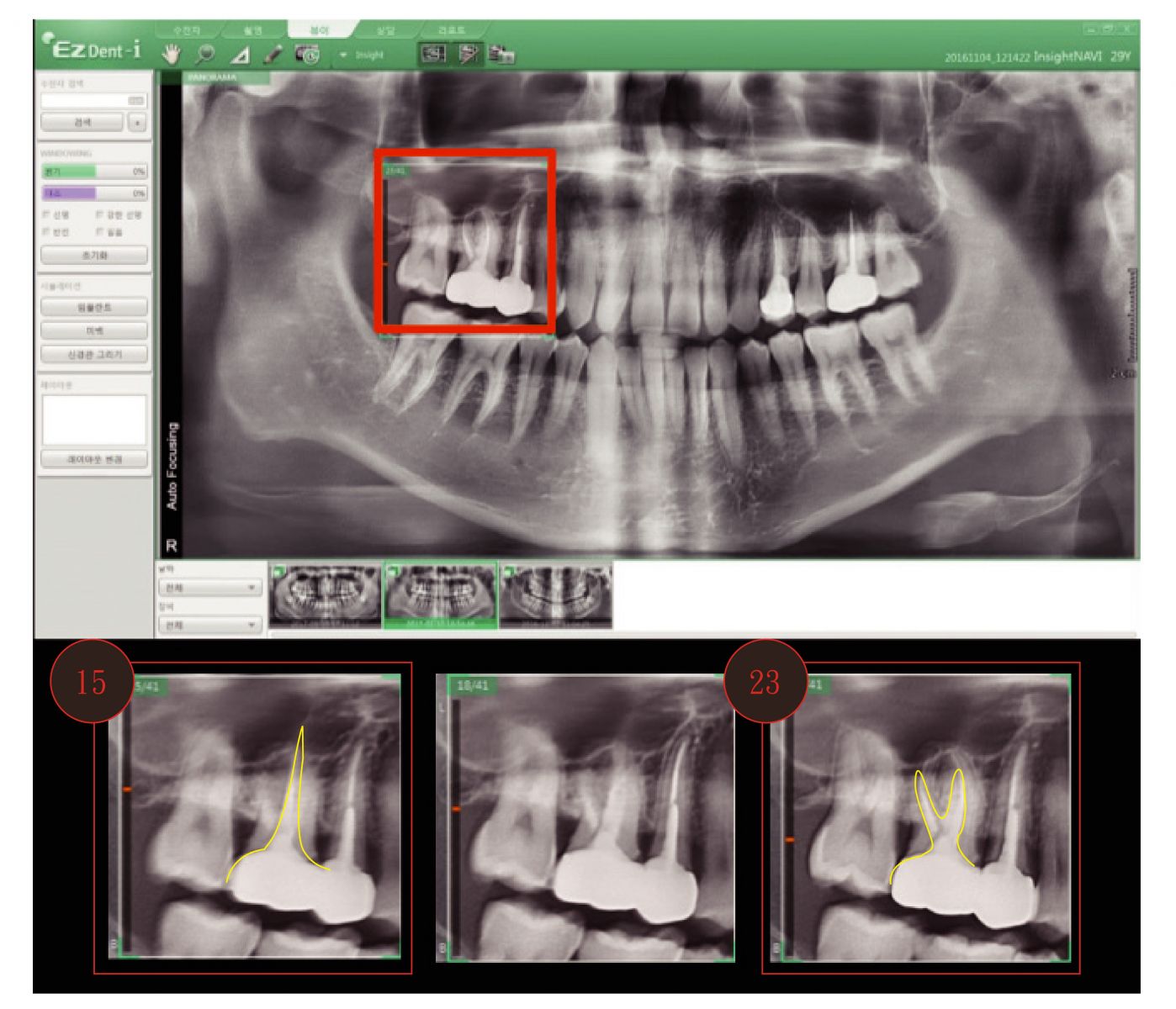
多牙根檢視 透過 41 剖面,檢視頰側牙根第 15 張剖面,顎側牙根第 23 張剖面,牙根組織狀況一覽無疑。 |
 |
PaX-i Insight has brought more pleasure and energy to work clinic and patients.
We use PaX-i Insight almost every day to get more detailed information for the accurate diagnosis and treatment. I can get all the depth information from 41 layered images, and this is exactly what a doctor needs for the completeness of work. The more the doctor has information about patient's condition, the more precise the diagnosis and treatment will be. PaX-i Insight would help doctors to find lesions and the information of which they are lack.
Nowadays, most of the pictures we capture are with Insight mode. And I wouldn't go back to the previous machine. If I have another chance to buy panorama X-ray, I would choose PaX-i Insight again which is the best choice in the market. With my experience with PaX-i Insight and VATECH, I would definitely recommend PaX-i Insight.
This is the exact case showing PaX-i Insight's value.
Loot at the sixteenth tooth. If you pass through the layers in the maxillary sinus, it is absolutely clear what the cause of this cyst was.
The gutta-purcha is overextended. It is even bent inside as a shape of the letter 'S'. This is very clearly visible if we are passing through the layers. The overextension of gutta-percha from the canal extending in-to the sinus and hidden caries. The completeness of the information is perfect, the image is clear and understandable. It helps us to make the prognosis of the patient. The ideal machine.
.jpg)
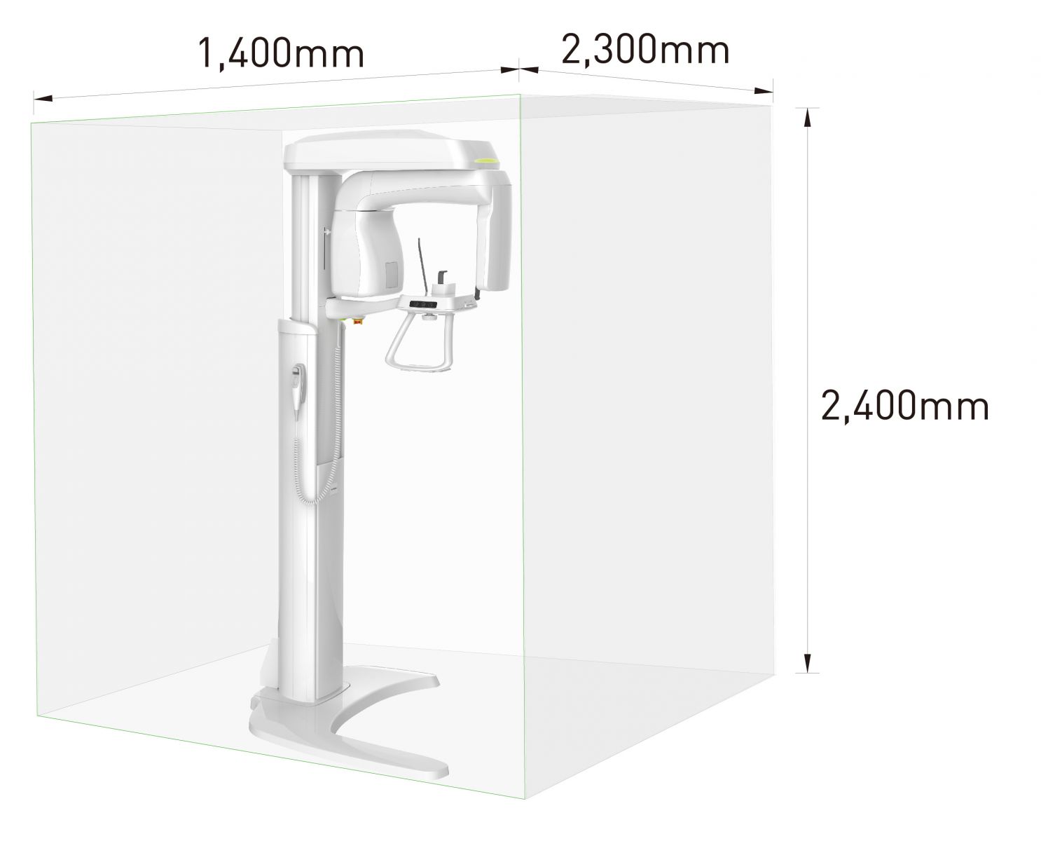
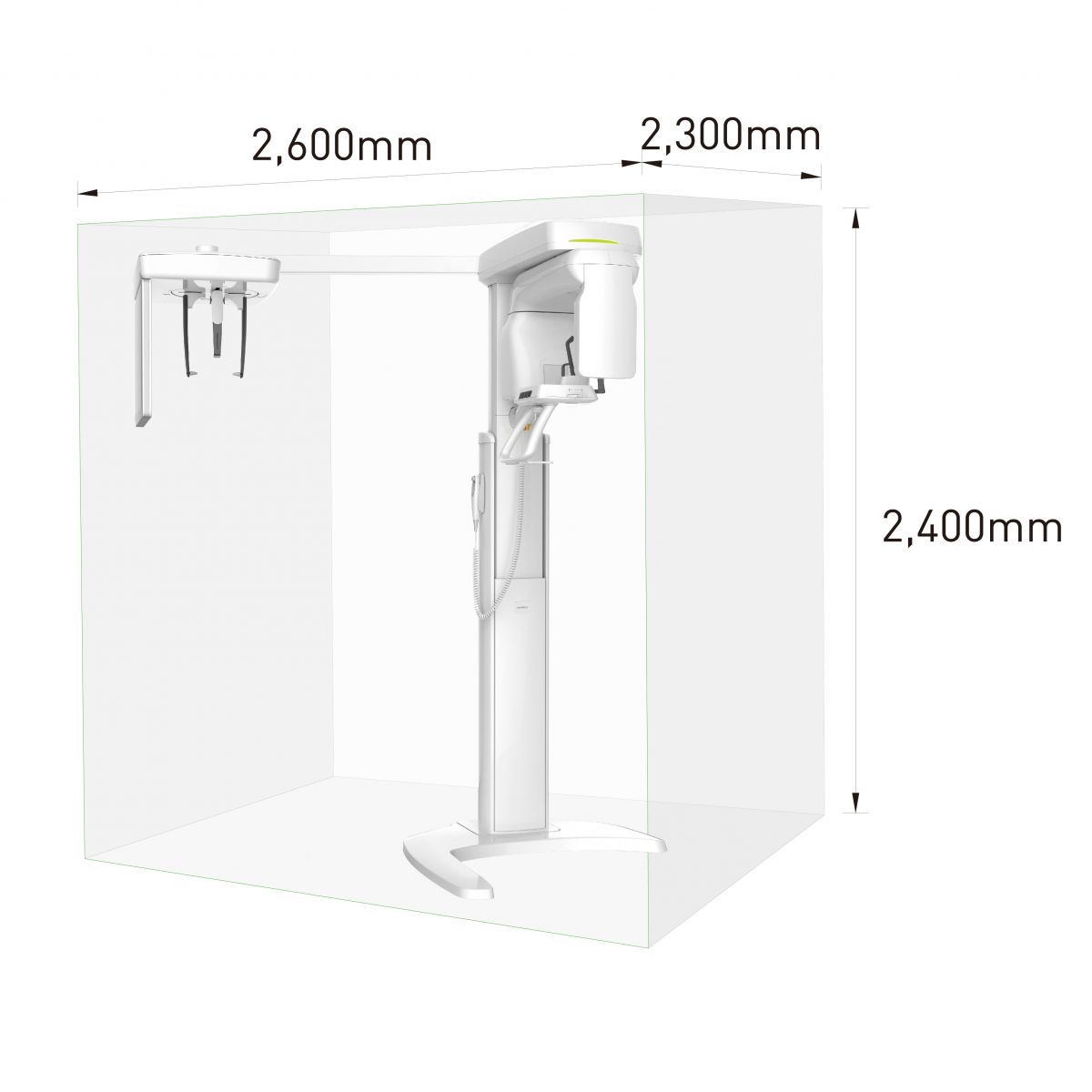
Dimensions
|
|
|
 |
 |
|
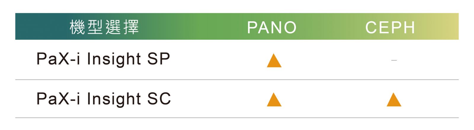
Function |
3in1 |
|
|
Function |
Pano |
• |
|
Ceph |
• |
|
|
Scan Time |
Pano |
10.4 / 14 |
|
Ceph |
3.9 / 1.9 |
|
|
Reconstruction Time |
< 2 mins |
|
|
Focal Spot [Unit: mm] |
0.5 |
|
|
Tube Voltage [Unit: kVp] |
60 - 99 |
|
|
Current [Unit: mA] |
4 - 16 |
|
|
Dimensions [Unit: mm] |
W/O Ceph |
990(L) x 1200(W) x 2300(H) |
|
W Ceph |
1930(L) x 1200(W) x 2300(H) |
|